EYE AND SOME DISEASE SYMPTOMS
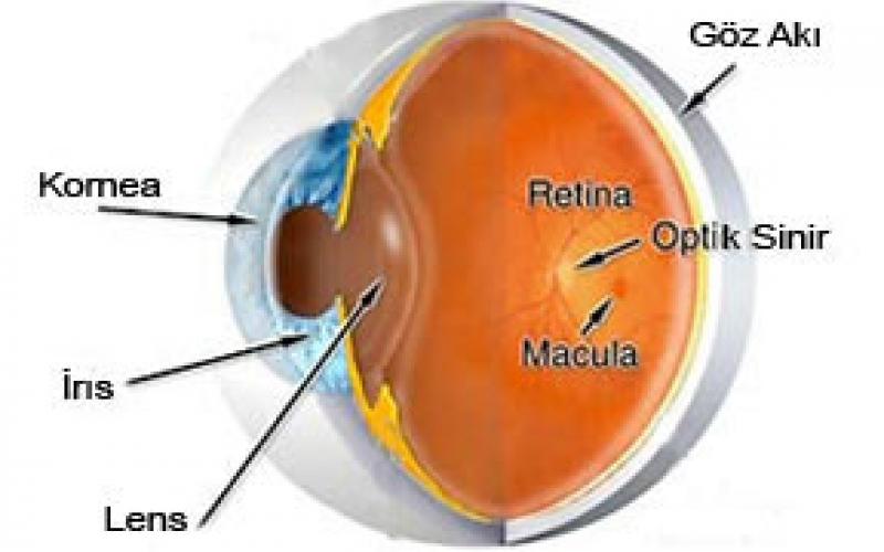
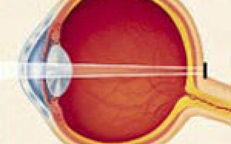
Eyeball: Eye is approximately 2.5 cm in diameter in the form of spheres. The outermost layer of the cornea (1) is in the middle of the white sclera layer in the form of a watch glass. The uvea constituting the middle layer consists of 3 parts: - the iris (4), which gives the color of the eye in front, - corpus ciare in the center, - and the choir, which allows most of the eyes to be fed. The most internal layer is the retina that marks the onset of the vision. A 2-ply transparent, slimy membrane called the conjunctiva surrounds the sclera. Between the corneal and the iris, there are chambers in the rear chamber, filled with aqueous humor between lens (intra-ocular lens) (3) and iris in the front chamber filled with transparent liquid (aqueous humor) with a depth of 2 mm between cornea and iris. The transparent vitreous behind the lens fills 3/4 of the eye, giving the shape of the sphere. At the back, there is an optical nerve that will transmit the view of the retinal to the brain.
Eyelids: It is the outer shell that protects the eye. The muscles in the covers structure allow periodic movement of the cover with the trimming reflex. The tear secreted from the tear gland acts as a wiper by the movement of the covers and prevents the layers in the front part of the eye from drying out and provides cleaning.
Cornea: It is the the frontmost, curved and veinless layer of the eye and the part where the contact lens is placed. It has approximately a diameter of 12 mm half a millimeter thick. It provides the greatest amount of cornea by breaking eye-catching light beams so that they form a clear picture of the retina (in the nerve layer). When the cornea refracts light ray a little, it causes hyperopia, when it refracts light rays much, it causes myopia and when it refracts light ray in equal directions, it causes astigmatism.
Iris: It is the vascular layer that gives the eye its color. Its contraction and relaxation with the muscles in its structure allows the pupil dilation and pupil constriction with a space in the middle. As it has constriction in the light and dilation in the dark, it balances the light rays entering the eye.
Lens: Behind the iris, it is a transparent veinless structure in 5 mm thickness and 9 mm diameter. Its function is the second layer that refracts the light rays entering the eye after the cornea. Its difference from the cornea is that the lens has the flexibility and refractiveness of the lens, that is to say, its zoom ability, so that we can clearly see the object at any distance .
Vitreous: It is a gel-like substance that fills the whole eye cavity behind the lens.
Retina: It is the nerve layer that covers the inner wall of the eye. The light rays entering the eye are transformed into electrical signals and transmitted to the eye nerve. Optic nerve (eye nerve) meets the nerve fibers from the retinal layer at one point of the eye and continues towards up to the brain.
HOW DO WE SEE?
The rays reflected from the objects we look at are primarily refracted by the transparent layer (Cornea) and the lens (Lens) in front of the eye and focus on the “Retina” layer consisting of nerve fibers located at the back of the eye. The image of the object formed in the retina is transported to the visual center of the brain via the optic nerve and vision occurs.
Astigmatism:
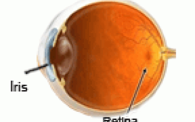
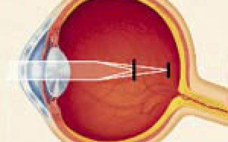
It is one of the refractive defects. It affects the focus of light in the retina, which disrupts the interpretation of the image in the brain. Astigmatism is usually caused by the curvature of the cornea having a slight egg shape. It is not a disease and is a very common problem. Blurred vision occurs in both far and near objects, which leads to eye fatigue, cross-eye and discomfort in individuals. Sometimes astigmatism occurs with myopia and hyperopia. These diseases called Myopic astigmatism and Hyperopia astigmatism can be treated with glasses and contact lenses.
Astigmatism
Myopia:
The person sees nearby objects clearly and sees far object as blurred, and the anterior posterior length of the eye is more than normal or the curvature of the cornea is too vertical. Images focus on the front side of the yellow point. Myopia usually begins at the age of 8-12 under 20 years. It may progress rapidly in adolescence. Correction is made with glasses and contact lenses.
Myopia
Hyperopia:
The person sees nearby objects clearly and sees far object as blurred. After close work, blurred vision, discomfort and fatigue develop in individuals. In careless examinations, hidden hyperopia may not be noticed. In the hyperopia eye, the eyeball is small or the cornea is very flat. Images focus behind the eye.
Hyperopia
Cataract:
It is the loss of transparency that develops in eye's own lens. It causes foggy-blurred vision. The incidence increases over the age of 55. Ultraviolet rays and cigarette use are thought to accelerate the development of cataracts, although there is no definite way to prevent it. Cataract seriously disrupts vision in advanced stages.
Lid Sebaceous Cyst:
Eyelids are swelling that can be confusable with hordeolum. It is an inflammation of the small oil-producing glands in the upper and lower eyelids. This occurs by blocking the glands. It may decrease and disappear by itself, hot compress application, drug injection into the swelling and sometimes surgery may be required. Frequent recurrent chalazions may be a sign of other diseases.
Conjunctivitis:
It is the inflammation of the thin membrane covering the white part of the eye. Bacterial infections, viral infections, allergies, environmental pollution (smoke and chemical vapors) may cause this condition. With increased blood flow in the conjunctiva, the eye looks red. It can be acute (intense), chronic (long-term). Burring and adherence in the eyelashes in the mornings may occur .
Allergic Conjunctivitis:
It is common in people with hay fever or seasonal allergies. Usually symptoms increase in spring. Red itchy eye and watering occur. Animal hairs, dust, pollen are the most common causes. Although it is easy to treat, it can sometimes be annoying.
Dry Eye:
Tears are very important for eye health. Contains natural lubricants & preservatives of eye Dry eyes may be in question in people with persistent eye stinging. The symptom of the dry eye can sometimes be paradoxically watering. This occurs as a reflex against stinging. Causes of dry eye are reduction in blink reflex, antihistaminic drugs, environmental factors (low humidity and wind), chemical and thermal burns, and arthritis. Artificial tear drops and gels are sometimes used for the treatment of artificial obstruction of lacrimal ducts Room humidifiers may be useful in dry areas.
Glaucoma (Eye Pressure):
Once the eye's own fluid is produced, it leaves the eye on the spongy membrane between the cornea and the sclera. A blockage in this region leads to increased eye pressure. Symptoms vary according to glaucoma type.Glaucoma occurs in 2 out of every 100 people over 35 years of age. People with glaucoma or myopia in their families have a higher risk. Early diagnosis with regular eye examinations is very important as the disease cannot be prevented and the damage to the optic nerve cannot be reversed. There are different types of glaucoma. However, they all result in damage to the optic nerve.
Chronic Open Angle Glaucoma:
It is the most common type. It is asymptomatic in people over 40 years of age. It usually causes irreparable harm to the patient before diagnosis. Therefore, regular eye pressure measurement is quite important.
Normotensive Glaucoma:
Although intraocular pressure is within normal limits, it is glaucoma due to impaired feeding of the optic disc. In people with thin corneas, the actual intraocular pressure measured is greater than the actual value in the eye. In this case, the eye nerve is affected. Besides, in some people with normal eye pressure (in the elderly), normotensive glaucoma occurs due to lack of blood intake of the optic nerve.
Congenital Glaucoma:
It occurs immediately after birth. There is a problem with the drainage system of the eye fluid. Babies with photosensitive or permanently irrigated eyes should be examined especially for this condition.
Acute Angle-Closure Glaucoma:
It occurs by sudden and complete closure of the eye fluid discharge area. The patient sees halo around the lights; there is severe pain, nausea and blurred vision. It may occur at any age. It requires emergency treatment Blindness may develop within 1-2 days.
Secondary Glaucoma:
It is a type of glaucoma occurring due to another eye problem. For example, glaucoma occurring due to uveitis.
Macular Degeneration
The macula is located in the center of the retina layer and is responsible for central vision. Central vision is impaired in this disease. There are two types. Dry type and wet type. The incidence increases over the age of 55. It is therefore called age-related macular degeneration.
Dry Type - Macular Degeneration:
It is seen at the rate of 90%. Patients see a black spot in the center or describe refraction on thin and long objects. It shows slow progress within years.
Age Type - Macular Degeneration:
New vessel formation is in question in the macula, which leads to easy bleeding and liquid leakage. Central vision can be lost quickly. With the help of FFA / ICG technique, new vessels can be detected and controlled by Argon Laser or photodynamic therapy.
Floating Specks:
Sometimes people may see spots and filamentous objects in the area they look at. Even though it can be seen at any age, it is more common in advanced ages. The inside of the eye is normally filled with a gel-like substance called the vitreous. Congenital protein residues in this material, water voids and spots in the visual field can be observed. In general, although floating specks can be a normal condition, they may indicate an eye disease.
(Uveitis, bleeding in the eye, retinal detachment, etc.)
Hordeolum:
It occurs by the formation of red swelling on the lids. It is inflammation of the eyelash follicle. Hot compresses and antibiotic ointments are used for treatment.
Blepharitis:
It is a chronic and stubborn eyelid inflammation that is frequently encountered in oily skin, dandruff and dry eyes. There is discomfort and itching in the eyes and dandruff and slight redness on the lips. Although there is no definite treatment, control can be provided with the drugs prescribed by the ophthalmologist.
VISION DISORDERS AND TREATMENT
Normal Eye
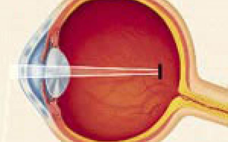
If the refracted rays in the cornea and lens fall exactly on the retina to the visual center, there is no refractive error and a clear vision is obtained.
Myopia (Shortsightedness)
The rays should focus on the retina; however, they focus on the front of the retina. With the refracted rays in the cornea and lens focusing relatively on the retina before reaching the visual center, the eye cannot see distant objects clearly. This condition is called Myopia.
Hyperopia (Farsightedness)
The rays cannot be refracted enough to focus on the retina. As the rays refracted in the cornea and lens do not focus relatively on the visual center on the retina, they focus on furhter area, the eye cannot see clearly. This condition is defined as Hypermetropia.
Astigmatism
The rays focus on more than one point in the retina. For a clear view, the cornea must be smooth and have the same curvature on each axis. If the cornea is more or less curved on a certain axis, it causes astigmatism defect. The image is not clear at both near and far visual spaces, and one sees objects as shaded.
MISUNDERSTANDINGS
- Reading in the dim light exhausts your eyes, but does not destroy it!
- Bright light disturbs the eyes, but does not destroy it. Sunglasses are not necessarily required!
- Excessive use of eyes exhausts them, but does not destroy!
- Strong, weak or wrong glasses exhaust the eyes, but do not destroy!
- Contact lenses and eyeglasses correct the eye disorder as long as they are worn; however, never completely eliminate the refractive errors! - In normal people, tears clean eyes sufficiently, no wetting eye drops are required!
- Headaches are not usually caused by eye fatigue, especially migraine at all! (Some headaches, such as ophthalmic migraine, may be triggered by light, etc., but not triggered by eye fatigue!)
- Healthy eyes do not require annual examination until the age of 35! (Of course, firstly, they must be checked if they are healthy!! )
- The eye with open angle glaucoma looks completely normal from the outside! (Eye with open angle glaucoma can only be understood by ocular blood pressure measurement and other examination methods...)
- Continuous watering of infants' eyes is caused by congenital glaucoma (congenital eye pressure) or obstruction of the lacrimal duct.
- Chemical substances entering to the eyes should be removed by washing with water without waiting.

