Excimer Laser
WHAT IS EXCIMER LASER?
It is a UV laser with a wavelength of 193 nm, obtained by electrically stimulating the argon-fluoride gas mixture. It has been used for the treatment of refractive errors for more than 25 years. It has an unbelievable sensitivity.
WHO CAN BENEFIT FROM THE LASER TREATMENT?
Myope persons up to 12 degrees, hyperopia persons up to 6 degrees and astigmatic persons up to 6 degrees between 18-60 years of age, not having some systemic diseases (diabetes - rheumatism, etc.) and chronic eye disease can benefit from this treatment.
HOW TO UNDERSTAND IF MY EYES ARE SUITABLE?
Detailed Optalmological Examination: Your visual acuity, detection of eye impairment, eye pressure measurement, biomicroscopic examination are performed. Then the pupils are enlarged with a drop and the eye numbers are re-determined by the drop and a detailed examination of the eye ground (retinal vessel structure and eye nerves) is carried out .
As the pupils will be enlarged by drop during the examination, there may be discomfort from light and blurred vision and may last for 24 hours. Automobile users should take this into account.
Examinations: In addition to detailed eye examination, corneal topography, pachymetry, Epithelial Thickness Map and pupillometer are carried out. The shape and thickness of the corneal layer are evaluated with these examinations.
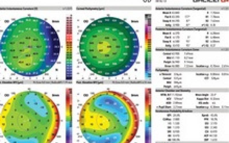
Topography: Topolaser, Orbscan and Gallei tests are used to determine the topographic map of the corneal layer of the eye in detail.
Corneal Topography Image
Gallei Topography Image
Pachymetry: Corneal thickness is measured. Corneal thickness is quite important for determining the surgical method to be applied.
Pupillometer: It is the process of measuring the pupil's diameter. The pupil becomes smaller and larger according to light intensity, and in some people the pupil is structurally larger. In particular, the planning of treatment in these persons according to this measurement is intended to prevent undesirable conditions such as post-laser light flare, image scattering, night vision problems and halo.
WHAT SHOULD I DO BEFORE COMING TO THE EXAMINATION?
Those wearing soft lenses should stop wearing their lenses one week before the examination and those wearing gas-permeable semi-hard lenses should stop wearing their lenses three weeks before the examination.
EVERY EYE IS DIFFERENT EXCIMER LASER TREATMENT WITH INTRALASE THROUGH BLADE-FREE LASER TECHNOLOGY
How to Perform Excimer Laser Treatment with INTRALASE?
1. Step: Creation of cornea map (3-dimensional):
Like the fingerprints, the corneal layer is also personal. The corneal topography device quickly and safely identifies the characteristics of your eyes; three-dimensional map is formed. This is a detailed map of not only the outer surface of your eye, but also how your eye directs light.

2. Step: Corneal flap formation:
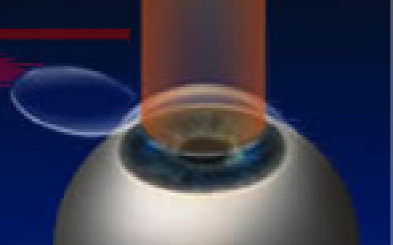
In the conventional method, a circular incision is made using a manual knife to form a corneal flap. Through the Intralase device, this process is performed with tiny, flawless laser shots, that is, completely without laser contact with your eye. When creating the flap, the 3D corneal map data created in the previous step is used; thus, the flap is created in the most appropriate position and in the most appropriate way to the structure of the eye.

3. Step: Wavefront-guided Excimer Laser treatment:
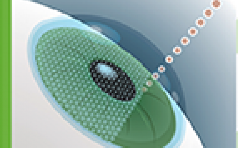
After the flap removal process performed by the intralase, the cornea is reshaped by applying laser process.
Why Intralase LASIK?
- It is safe because it is performed with completely computer controlled devices.
- It can also be applied to patients with thin corneas.
- It can be applied to patients with high visual impairment.
- It allows astigmatic correction.
- It minimizes the possibility of corneal flap complication.
- Treatment is completed in minutes.
- It is a painless and indolent procedure.
- Most patients notice improvement in vision immediately after treatment.
- Recovery is fast.
HOW TO APPLY LASIK (Lazer In-situ Keratomileusis)?
A thin layer (flap) of approximately 160 microns is removed from the cornea (transparent tissue at the front of the eye). The lower layer is reshaped with the help of Excimer Laser. Removal of this layer (flap) is important for the permanence of the correction and rapid recovery.
Flap Removal
Laser Application
Laser Application
WHAT HAPPENS AFTER THE OPERATION?
Burning, watering and blurred vision in the eyes are normal for 4–5 hours. Drops are given every 1-2 hours. After a 5-minute follow-up on the next day, patients can return to their daily work.
WHAT SHOULD I CONSIDER AFTER THE SURGERY?
On the next day of the operation, it is free to wash the face (keeping eyes closed) and take a bath without missing soap or shampoo to the eye. Do not enter the pool or sea for 3 weeks and do not make up for 2 weeks. Scrubbing and scratching the eyes should be avoided.
WHAT ARE THE ADVANTAGES OF T-CAT (Topolyser Customized Ablation Treatment)?
The laser makes a treatment plan according to the map that it reads in full detail. This plan also includes a part of the wavefront method. The treatment plan will be made by laser by evaluating both the map and wavefront results together.
The system determines the ideal shape of the corneal layer after treatment and transfers this shape to the laser with a special formula.
All data are transferred to the treatment mode of the laser with the consent of the physician and the treatment is administered. The administration stage of the treatment for the patient is no different from the standard lasik application. The difference is the preparation of the treatment and the shape of the laser on the cornea.
PRK and LASEK Method
In the PRK and Lasek methods, epithelial tissue outermost of the eye is removed before laser treatment and then laser treatment is performed. The PRK system is older than LASEK. In the PRK method, epithelial tissue is stripped with the help of a surgical instrument. In LASEK method, this tissue is removed in the form of a layer with the help of alcohol and put into its place after the laser treatment. The tissues heal within 3-4 days after the operation and combine the epithelial tissue with the upper part of the eye again. However, in some patients, this process may be painful. It may take 3 to 4 weeks for the eye to fully heal.
WHAT ARE ALTERNATIVE TREATMENT METHODS IN CASE THE PATIENT IS NOT SUITABLE FOR LASER TREATMENT?
For patients who cannot be treated with laser for various reasons, there are treatment alternatives such as phakic intraocular lens, ICL, Toric ICL, refractive lens replacement, RK, Corneal Cross Linking (CCL) and INTACS.
Toric ICL
Phakic Intraocular Lens
ICL (Implantable Contact Lens) – TORIC ICL
ICL and Toric ICL can be used in cases where laser treatment cannot be performed. For example; ICL is applied to moderate to high myopia and hyperopia eyes, while Toric ICL is applied to myopic astigmatism eyes. It is a preferred treatment method in people with thin corneal layer and dry eye.
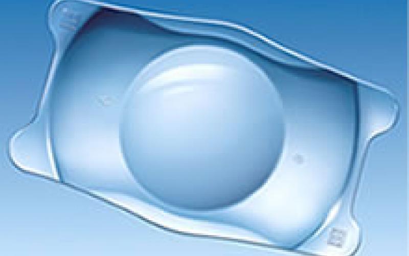
Since the toric ICL is small and soft, a small incision can be made in the cornea in the anterior part of the eye and folded to be injected into the eye without pain within a few seconds. After injection, the Toric ICL has the required position in the fluid between the iris and the natural lens. The lens is easily accepted by the body.
PHACIC IOL
Refractivce error in patients who has too high values to be corrected by Excimer Laser (myopia values 8 to 25 and hyperopia values 6 to 13) and in patients who has thin cornea is corrected by placing a numbered artificial lens into the eye. The artificial lens placed is transparent, remains in the eye for life, is not felt by the patient and is not visible from the outside. The operation time is 15–20 minutes and is not performed on both eyes at the same time. The operation is usually performed under general anesthesia in younger patients and later under local anesthesia. The eye is closed 1 day after the operation. The next day, the patient begins to see clearly.
CORNEAL CROSSLINKING (Molecular Crosslinking)
This treatment was implemented by Prof. Theo Seiler and his team in Switzerland for first time in the world. It is thought that UV - Cross Linking operation can effect the collagen molecules of the cornea by using UVA light and Riboflavin and increase the corneal mechanics and halt the progression of keratoconus disease. The studies carried out have revealed that the progression of the disease is stopped and the corneal curvature is improved by 2d. No side effects were detected in the studies.
How to apply CORNEAL CROSS LINKING (Molecular Cross Linking) treatment?
In order to apply this treatment, it is important to determine the fact that the disease is progressive. As a result of detailed examinations, it is revealed that your disease is progressive and then treatment is applied to the appropriate cases. The eye is anesthetized with topical anesthetic drops before the procedure. Following topical anesthetic drops, the corneal epithelium is removed with a blunt spatula. Riboflavin solution is dropped to the cornea with epithelium removed for 30 minutes with 2 drops at 5 minutes internals. After 30 minutes, the patient is placed on a biomicroscope. After riboflavin fluorescence is seen in the anterior chamber, the patient is taken back to the operation room. 370 nm UV is applied for 30 minutes in an area of approximately 7 mm at a distance of 4-5 cm from the corneal surface. During the UV application, 2 drops of Riboflavin are dropped every 5 minutes. After the procedure, protective contact lens is applied to the eye and only closure can be applied in cases deemed necessary.
INTACS
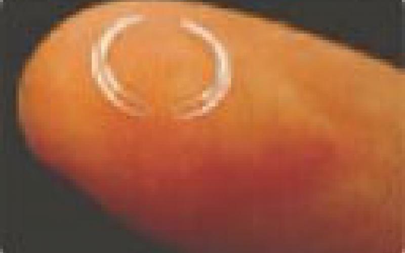
Intacs operation is performed for keratoconus patients and for the treatment of corneal complications occurring after failed or incorrect laser surgery. The corneal rings used are made of synthetic material and are round rings placed inside and close to the outer edge of the cornea. With the special surgical set of the Intacs device used in this operation, firtst the channels where the rings are placed are created. The rings are then inserted into the cornea. The rings are designed to form a 150 degree circle when placed. The rings press the circumference of the cornea and act by flattening this area.
